Blood staining techniques like Wright-Giemsa Stain are vital tools in clinical diagnostics. Today, these tests play an indispensable role in the identification and analysis of blood cells and microorganisms. These techniques allow laboratory professionals to:
- Distinguish between various cell types
- Detect abnormalities
- Identify pathogens
The blood smear stain continues to be a critical step in routine hematology and infectious disease diagnostics. Therefore, the various types of staining methods make it easier for effective diagnosis and treatment strategies as they provide detailed cellular imagery.
However, despite their importance, traditional stains such as the Wright-Giemsa stain have several limitations. Examples include time-consuming procedures, high operational expenses, dependency on liquid reagents, and environmental burdens.
These challenges shed some light on the urgent need for more sustainable, efficient, and scalable staining technologies, such as the Next-Generation Staining and Immunostaining (NGSI) solution developed by NOUL. The FDA-listed fluorescence imaging developments strengthen the potential of NOUL’s approach to diagnostics.
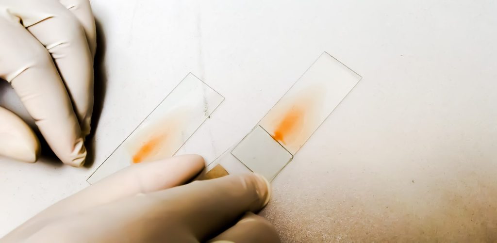
Wright-Giemsa Stain: A Widely Used Method
Source: Shutterstock
The Wright-Giemsa stain is a commonly used variant within the Romanowsky stain family, combining features of both Wright and Giemsa stains to provide enhanced cellular contrast and morphological detail in peripheral blood and bone marrow smears. This stain combines eosin Y (an acidic dye) and methylene blue (a basic dye). These substances bind to cellular components based on their chemical affinity. As a result, red blood cells, white blood cells, and platelets can be differentiated clearly under a microscope.
The staining mechanism is based on the Romanowsky effect, in which a combination of acidic (eosin Y) and basic (methylene blue) dyes interact with cellular components to produce sharp contrast. This effect enables nuclei to appear purple, cytoplasm to vary from pink to blue, and granules in certain white blood cells to be distinctly colored. As such, it allows detailed morphological differentiation that is critical in hematological assessments.
Due to its low cost and availability, the Wright-Giemsa stain is widely used in hematology labs to diagnose infections such as malaria and various blood disorders. The main advantages lie in its cost-effectiveness, simplicity, and adaptability in resource-limited environments. Moreover, both Giemsa and Wright-Giemsa stain procedures remain cornerstones in the detection of blood parasites and cytological studies.
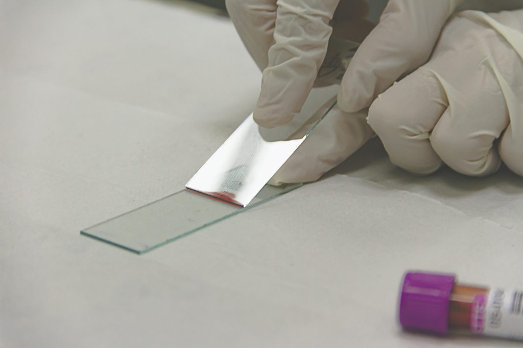
Romanowsky Stains: The Broader Family Behind Wright-Giemsa
Source: Pexels
The Romanowsky stain family includes several stains, such as Wright, Leishman, and Giemsa stain procedures. These methods are known for their capacity to produce contrasting images of blood cells, which are vital to identify abnormalities and infections. Specifically, many developing regions prefer Romanowsky stain techniques due to their reliability and ease of use.
Originally developed by Dmitri Romanowsky in 1891, this class of stains was a breakthrough in cytological visualization. The hallmark Romanowsky effect, resulting from the interaction of azure B (a methylene blue oxidation product) and eosin, gives rise to the distinctive purple coloration of chromatin. This innovation formed the backbone of modern blood smear techniques, and its simplicity and clarity remain unparalleled, especially in low-resource settings.
They require only basic laboratory setups and minimal technical training, which makes them ideal for routine use in clinical labs. The blood smear stain created by Romanowsky-type staining helps healthcare providers to clearly see the shape of cells and count different types of white blood cells, which is important for diagnosing leukemia, anemia, and infections.
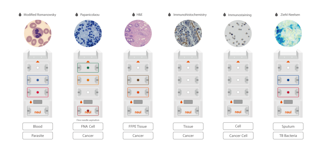
NGSI Technology by NOUL: Addressing Limitations
Source: NOUL
Although the Wright-Giemsa stain and other types of staining within the Romanowsky stain category serve well in many diagnostic scenarios, they require:
- Liquid reagents
- Precise timing
- Extensive washing
- Waste disposal systems
These factors limit their scalability and pose safety and environmental risks.
NOUL’s NGSI (Next-Generation Staining and Immunostaining) technology presents a revolutionary alternative. NGSI uses hydrogel-based patches that can be embedded with eosin and methylene blue, which are pressed directly onto the blood smear. This method eliminates the need for immersion in staining solutions. The solid-based staining technology simplifies the process, minimizes user error, and eliminates wastewater generation.
The benefits of NGSI include reduced reagent use, safer handling, and decreased hazardous waste generation. Its compatibility with NOUL’s miLab™ platform supports portable diagnostics, especially in under-resourced settings such as field hospitals and rural clinics. NGSI’s compact format and reproducibility align with the global need for scalable diagnostic technologies, especially in regions that face limited healthcare infrastructure.
For a technical breakdown of the method, NOUL has published details on their hydrogel-based NGSI technology, which shows the scientific innovation behind their solution.
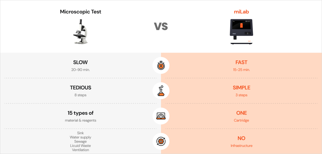
The following table summarizes the main differences between traditional straining methods and NGSI technology:
Conclusion
Source: NOUL
Conventional types of staining methods, such as the Romanowsky and Wright-Giemsa stains, have long supported hematological and infectious disease diagnostics. However, the procedural intricacies, waste management issues, and limited automation potential create barriers, especially in resource-poor environments.
With the introduction of NGSI technology and platforms such as miLab™, NOUL has set a new standard in the world of blood smear stain techniques. This next-generation solution eliminates liquid reagents and reduces staining steps. The goal is to allow laboratories to provide faster, cleaner, and more accurate diagnostics.
Healthcare professionals who are interested in the diagnostic capabilities and sustainability of this technology can contact NOUL directly for partnership opportunities.