Whenever you receive a cut, your blood clots to seal the wound, forming a barrier against infection, reducing blood loss, and ensuring your safety.
Platelets, or thrombocytes, are small, disc-shaped cells in the blood that enable clotting. When a blood vessel is damaged, platelets activate at this site, releasing chemicals that help them stick together and form a clot.
Disorders arise when the number or size of platelets differs from normal. The most common condition is thrombocytosis, an abnormally high platelet count. Medical professionals use several metrics to monitor platelet health:
- Platelet Count: The total number of platelets.
- Mean platelet volume (MPV): The average size of platelets.
- Platelet Distribution Width (PDW): The variation in platelet size.
These metrics aid in diagnosing conditions such as thrombocytosis, giant platelets, and abnormal platelet clumping. Let’s explore these in detail.
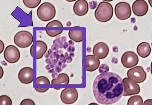
Thrombocytosis: Definition and Causes
 Source : Shutterstock
Thrombocytosis refers to an abnormally high platelet count, classified into two types:
- Primary Thrombocytosis (or Essential Thrombocytosis): Caused by bone marrow abnormalities, often linked to genetic mutations like JAK2.
- Secondary Thrombocytosis (or Reactive Thrombocytosis): A reaction to another condition, such as infection or inflammation, where chemical messengers temporarily increase platelet production.
Mean Platelet Volume (MPV): Assessing Platelet Function
Mean platelet volume (MPV) measures the average size of your platelets. Platelets must be the right size to fit through blood vessels and clot normally. The normal MPV is 7.5 to 12 femtoliters (fL).
If your MPV is too high, then there are lots of larger, more activated platelets in your bloodstream. It often occurs with thrombocytosis or marrow diseases. However, the most common cause is blood loss – the body makes extra platelets to compensate, which leads to a rise in the larger precursor megakaryocytes.
If your MPV is too low, it usually means that platelet production has slowed, and the remaining cells are much older. This occurs as a result of aplastic anemia, inflammation, or cancer.
MPV is compared to platelet count. Together, they can be diagnostic. For example, a high MPV and low platelet count occur in conditions like immune thrombocytopenic purpura, while a high platelet count increases the risk of a genetic mutation or cancer.
Platelet Distribution Width (PDW): The Importance of Platelet Variability

Source : Shutterstock
Platelet distribution width, or PDW, indicates the variability in platelet size. It’s a marker of platelet function and activation. A normal PDW ranges from 9 to 14fL. Usually, a normal PDW suggests that platelets in the bloodstream are similar sizes – a good sign.
However, if the PDW is too high, then the variation in size is much higher. It occurs due to thrombocytosis (if the platelet count is also high) or conditions like iron deficiency anemia, myeloproliferative disorders, chronic inflammation, or bone marrow disorders.
Low PDW is generally considered healthy. However, it can also occur in conditions where there is minimal platelet production or abnormal platelet agglutination.
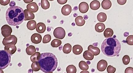
Giant Platelets: Characteristics and Diagnostic Significance

Source : Shutterstock
Giant platelets, or macrothrombocytopenia (MTC), are a rare blood disorder where platelets are abnormally large. People with the condition tend to have fewer and larger platelets, which often struggle to bind together or become stuck on the blood vessel walls.
Causes of Giant Platelets
Genetic conditions like Bernard-Soulier syndrome are the primary cause of giant platelets. Changes in the GP1BA, GP1BB, or GP9 genes can cause the syndrome, as a person needs only to inherit one changed copy of one of the genes to develop the condition.
Other genetic conditions include May-Hegglin anomaly, Gray platelet syndrome, and myelodysplastic syndrome.
Caring for people with giant platelets requires consistent platelet transfusions and antifibrinolytic agents to reduce the risk of excessive bleeding.
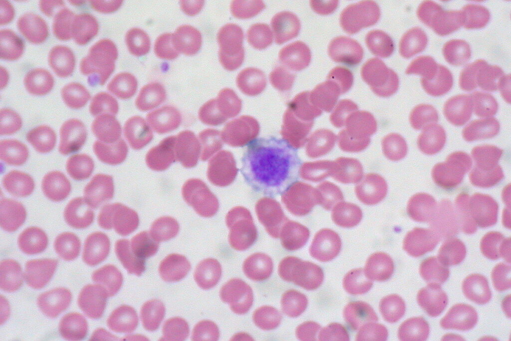
Agglutination: Understanding Platelet Clumping
 Source : NOUL
Agglutination is the technical term for the clumping together of platelets. It’s what’s supposed to happen during clotting. However, it can also occur due to infection, antibodies, or other immune responses. This happens when chemical messengers known as cytokines trigger an agglutination response.
If platelet agglutination occurs when a blood sample is taken, it can lead to an apparent decrease in the platelet count. Indeed, it might even lead to changes in the MPV, usually leading to a higher MPV.
Comprehensive Diagnosis: How Platelet Tests Work Together
 Source : NOUL
These different factors cannot be understood in isolation. Only by taking the test results together can medical professionals discern what’s going on. Platelet count is the most important factor, as it informs us whether thrombocytosis is occurring or not. However, interpreting this result requires both MPV and PDW.
For example, if your platelet count and MPV are low, it indicates a bone marrow disorder slowing down platelet production. Yet, if the platelet count is normal, it’s characteristic of chronic kidney failure. And a high platelet count and low MPV are indicative of an infection, inflammation, or cancer. Each constellation of results points to a different diagnosis; only together can you diagnose the problem.
Advancing Platelet Diagnostics for Better Care
 Source: Flickr